

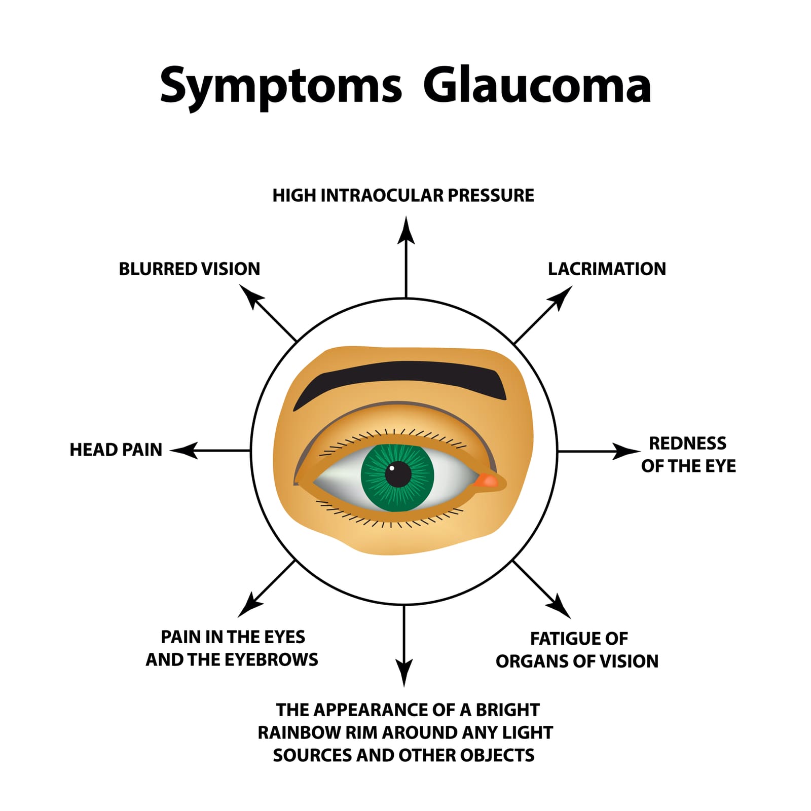
Glaucoma is a series of conditions that result from damage to the optic nerve, impacting vision. It is the leading cause of blindness worldwide. Examination of a glaucomatous optic nerve reveals "cupping," which looks like a "hollowing out" of the optic nerve head
How Glaucoma Affects Vision
Vision begins with the eyes. Light enters the eye through the pupil and hits the retina in the back of the eye. This trigger special vision cells called rods and cones to become activated in response to the light patterns. Information from the rods and cones is collected and passes into a single bundle called the optic nerve. The optic nerve is responsible for transmitting information from the eyes to the brain, where it will be processed.
The eye is filled with fluid. Fluid normally drains out through a special drainage system called the trabecular meshwork. When this drainage system doesn’t work right or too much fluid is produced, it can lead to a build-up of excess fluid. This causes the pressure within the eyeball to get too high. Excessive pressure leads to nerve damage within the optic nerve. As a result, people develop blind spots in their vision because visual information cannot get transmitted to the brain.
Types of Glaucoma
There are several types of glaucoma. All of them share features of optic nerve damage and resulting vision loss. The types of glaucoma include:
· Open-angle glaucoma. This is the most common form of glaucoma. Open-angle glaucoma is a leading cause of irreversible blindness in the world. It occurs when the drainage network is partially blocked, increasing pressure on the optic nerve. Vision changes occur slowly and gradually. Individuals with open-angle glaucoma rarely experience symptoms. Thus, open-angle glaucoma is generally detected incidentally during comprehensive ophthalmic examination. High elevations of intraocular pressure (IOP), up to 40 mmHg in patients with open-angle glaucoma, generally cause no pain, redness, or visual symptoms. There is no loss of visual acuity as long as central vision is preserved. Central visual field loss is a late manifestation of open-angle glaucoma, usually preceded by ganglion cell loss and optic nerve damage. Some patients are unaware of field loss even when it has progressed to central "tunnel vision" of 10 to 20 degrees. Visual field loss cannot be recovered once it has occurred. The mean progression rate from a full field of vision to blindness takes approximately 25 years in untreated patients.
· Angle-closure glaucoma. Also called closed-angle glaucoma, this type of glaucoma occurs when the iris of the eye bulges. This prevents fluid from circulating through the eye, increasing pressure. Angle-closure glaucoma can begin suddenly, causing rapid vision loss. It is a medical emergency that requires immediate treatment to prevent blindness.
· Normal-tension glaucoma. In this form of glaucoma, eye pressure stays within the normal range, yet the optic nerve gets damaged. This may be due to the build-up of fatty deposits within blood vessels serving the optic nerve.
· Pigmentary glaucoma. The iris, or colored part of your eye, contains pigment granules that give your eye its color. If these granules get dislodged, they can block fluid from exiting the eye.
· Childhood glaucoma. Glaucoma most often affects older adults, but children can develop it too. This may be due to an underlying medical condition or blockages of the drainage system.
Risk Factors for Glaucoma
Certain people are at higher risk for developing glaucoma. Major risk factors include
· Being older than 60 years old
· Certain racial and ethnic backgrounds, including being black, Asian, or HIspanic
· Medical conditions like high blood pressure, heart disease, and diabetes
· Family history of glaucoma
· Eye conditions such as nearsightedness, farsightedness, or thin corneas
· Using corticosteroid medications (particularly eyedrops) for a long period of time
· Past eye injury or eye surgery
Glaucoma Symptoms
Open Angle Glaucoma is usually asymptomatic and generally detected incidentally during comprehensive ophthalmic examination
Angle-closure glaucoma happens when the fluid-draining system in the eye gets blocked. This causes eye pressure to rise quickly and is a medical emergency. The symptoms include:
●Vision loss
●Seeing halos around lights
●Headache
●Very bad eye pain
●Feeling sick (nausea) and throwing up (vomiting)
●Redness in the white part of the eye
●Swelling and cloudiness in the colored part of the eye
Diagnosis of Glaucoma
It is critical to schedule annual eye exams to monitor your risk for glaucoma. Everyone age 40 and older should be tested for open-angle glaucoma at least once. Detecting it early can help to slow the progression of the disease, possibly preserving your vision. When you go to the eye doctor, your doctor will review your medical history and perform a comprehensive eye examination. This might include:
· Testing your visual fields to detect areas of vision loss
· Dilating your eyes to view the optic nerve
· Using a small device to measure the thickness of the cornea (pachymetry)
· Using a special viewer to inspect the drainage system of your eye (gonioscopy)
· Measuring intraocular pressure, a process called tonometry. This involves gently placing a device at the surface of the eye, which is typically numbed with eye drops. Your doctor may also send a puff of air onto the eye’s surface. Elevated IOP alone does not establish the diagnosis of open-angle glaucoma. One-third to one-half of individuals with glaucoma field defects have intraocular pressures ≤21 mmHg when first detected (normal IOP 8 to 21 mmHg) In addition, over 90 percent of adults with pressures >21 mmHg have no optic nerve damage. However, patients with elevated IOP should be referred to an ophthalmologist given their higher risk of open-angle glaucoma. Patients with elevated IOP but normal optic nerves and visual fields may be diagnosed as having ocular hypertension.
· Optical coherence tomography – Optical coherence tomography (OCT), Heidelberg retinal tomography (HRT), and scanning laser polarimetry are noninvasive imaging techniques that analyze light reflected off the fundus. The devices generate a digital image and quantification of specific features of optic nerve head anatomy.
· Visual field testing with brain computer interface – A portable brain-computer interface (wearable, wireless, dry electroencephalogram and electrooculogram systems) that assesses electrical brain responses to visual field stimulation has shown promise as an alternative and potentially more reliable means of diagnosing glaucoma compared with standard visual field testing.
Glaucoma Treatment Options
Once you have damage to the optic nerve, it cannot be reversed. The goal of glaucoma treatment is to slow down the progression of the disease. With early detection and intervention, it is possible to prevent additional vision loss.
Eye Drops: The most common treatment for glaucoma is eye drop medication. Eye drops lower the pressure within your eyeball. Some drops lower the amount of fluid your eye produces, while others help fluid flow through the drainage angle more easily. Side effects of eye drops include a stinging sensation, red eyes, red skin around the eyes, blurred vision, and changes in your pulse. Medications include
The topical prostaglandins -include Latanoprost, Bimatoprost and tafluprost. Ocular side effects of prostaglandins include hyperemia, eye irritation, increase in the number and length of eyelashes, and changes in iris and lash pigmentation.
Topical Betablockers- Timolol and betaxolol. Topical beta blockers are contraindicated in some patients with pulmonary or cardiac disease.
Alpha adrenergic agonists (eg, Brimonidine) appear to be similarly effective to beta blockers in lowering IOP in open-angle glaucoma,
Topical carbonic anhydrase inhibitors e.g Dorzolamide
Drug Combinations – Varying combination of drugs above.
Laser Therapy
There are two types of laser surgery, one for open-angle glaucoma and the other for closed-angle glaucoma. Each of them focuses on increasing the efficiency of fluid drainage from the eye. Laser surgery is relatively low risk.
Surgery
Surgical therapy (trabeculectomy) involves the creation of a filtration bleb to allow egress of aqueous humor from the eye. This is an alternative route to the normal pathway via the trabecular meshwork. Glaucoma surgery may sometimes involve the placement of mechanical shunts/drainage tube (also called "valves”) in patients who do not respond to other treatment.
Talk to your ophthalmologist to determine what treatment is best for you.
Medication Warnings in Glaucoma
Patients who are known to have narrow angles but who have not yet undergone laser peripheral iridotomy should avoid medicines that induce pupillary dilation and might provoke an angle-closure attack. Such medicines include over-the-counter decongestants, motion sickness medications, adrenergic agents, antipsychotics, antidepressants, and anticholinergic agents.
Steroids/Glucocorticoids can raise intraocular pressure in open angle glaucoma.
Sources
General information, not medical advice
This article can't account for your health history or current treatment. Talk to a qualified clinician before you change anything about your care.
Read our medical disclaimer →
On this page
How Glaucoma Affects Vision
Types of Glaucoma
Risk Factors for Glaucoma
Certain people are at higher risk for developing glaucoma. Major risk factors include
Glaucoma Symptoms
Diagnosis of Glaucoma
Glaucoma Treatment Options
Laser Therapy
Surgery
Medication Warnings in Glaucoma
Sources
Put what you read to work
Track your medicines, check a symptom and talk with people managing the same condition, all in Healthread.
Get the app
Keep reading

Beauty and Anti-Aging
Gastric Balloon for Weight Loss
The gastric balloon weight loss procedure is a new FDA-approved minimally invasive bariatric procedure for treating obesity. It is a temporary procedure that involves placing a saline-filled silicone balloon in the…
June 19, 2025 · 3 min read

Beauty and Anti-Aging
Vitamin D Deficiency
Also known as the sunshine vitamin, vitamin D is an essential nutrient that is naturally absorbed by the skin through exposure to sunlight. It can also be obtained from dietary sources and supplements.
December 30, 2024 · 3 min read

Cancer-Updates
Gynecologic Cancer
Gynecologic cancer is a term used to describe a group of cancers that affect the female reproductive system. There are five types of gynecologic cancers named after the body part where they start.
November 5, 2024 · 3 min read


© 2026 Healthread
English · Español
Healthread does not provide medical advice, diagnosis or treatment. Always speak to a qualified clinician about your care.